Crédits:
meteorites11111 le 18 juil. 2011
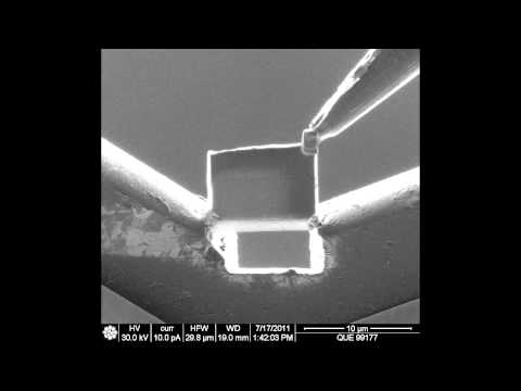
FIB lift out of CR 3.0 meteorite QUE 99177
The images are captured in a Scanning Electron Microscope, so they are pictures of backscattered electrons, with different contrast showing different chemistry. The surface where I extract a thin little biscuit of science is a piece of a meteorite. The Focused Ion Beam is a beam of big fat Gallium ions that bombard my sample.
First, I build a layer of platinum metal on the surface where I plan to make a sample section. This layer protects the sensitive meteorite sample from the ion beam. The platinum is deposited by cracking huge molecules of gas that is laced with platinum metal. When the molecules crack open, metal splatters down onto the surface directly below. So I can basically draw a shape and have it fill up with platinum.
Next, the holes are excavated using the focused ion beam. The big ions in the beam essentially act as microscopic wrecking balls, wreaking havoc and digging up a hole wherever I point them. So I dig holes on both sides of the sample section I am creating. Then I come in with a really tiny needle and adhere the sample to the needle. Then I free the sample from the meteorite and it zooms off with the needle.
Next, I adhere the sample to a sample grid using more platinum. Then I tilt the sample grid into the ion beam and shave off very tiny amounts of meteorite until it is ultra thin (~80 nanometers is the goal). The point is to create a sample that is so thin that I can transmit a beam of electrons through it in a transmission electron microscope. This allows me to view the atomic structure and chemistry of the meteorite, thus giving me an idea of how and where the meteorite bits formed and under what sorts of conditions.